Chest wall lumps
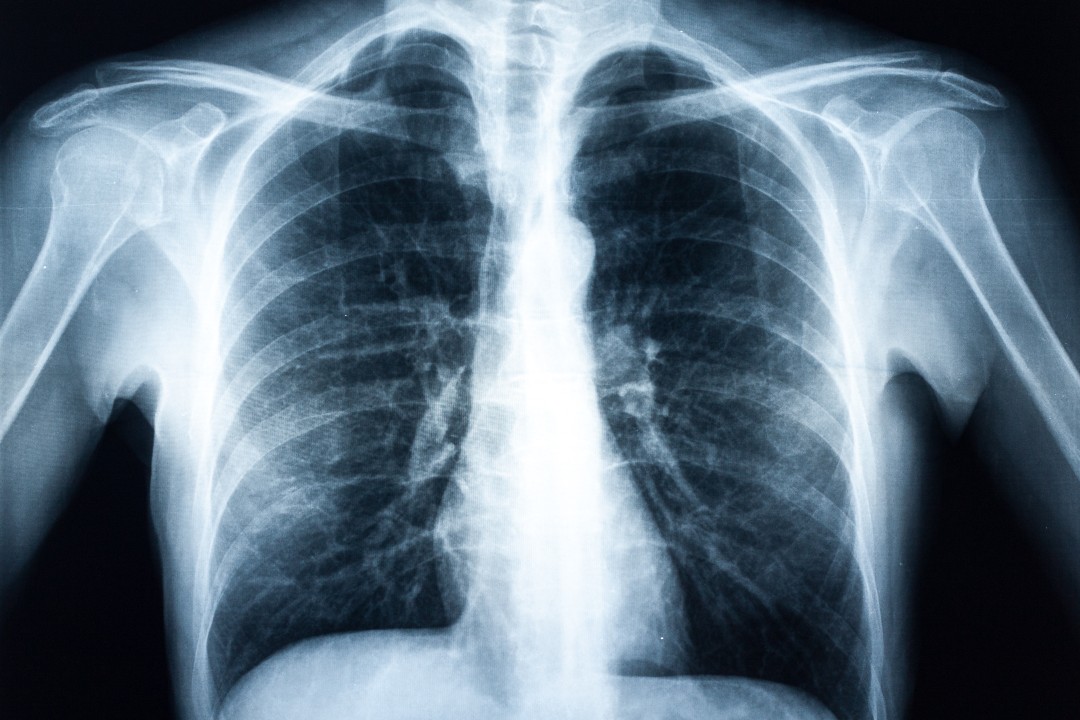
Chest wall injuries, inflammatory or infective conditions and even cancerous or non-cancerous growths can lead to the development of a chest wall lump. The chest wall is made up of bone (sternum, ribs and thoracic spine), cartilage and other ‘soft tissues’ including muscles, tendons, ligaments, fascia (membranes), blood vessels, nerves, lymph vessels and nodes. Any of these can lead to the development of a chest wall lump. In addition, underlying organs such as the lung, the lining of the lung (the pleura) or conditions in the cavity of the chest (the pleural cavity) can also lead to chest wall lumps.
What are the symptoms?
Depends on what the chest wall lump is caused by, but along with the lump or deformity there may be associated pain, swelling, inflammation (redness), discharge, clicking or altered sensation. If large they can cause a restriction of certain movements. The lump may quickly develop such as an abscess, or slowly or become noticeable with significant weight loss or with certain activities for example a lung hernia.
How is it diagnosed?
Having discussed how your chest symptoms developed and having carefully examined your chest, the specialist’s diagnosis is usually backed up by an ultrasound, chest MRI or a CT scan to show the lump depending on what the lump is caused by.
How is it treated?
Treatment is very much dependent on what the lump is caused by:
Non-operative treatment: It may involve nothing more than reassurance, rest or medication to treat condition.
Surgery: If required the lump may be removed.
How long does it take to recover?
Recovery will depend on the type of injury and treatment you’ve had. Your specialist will be able to advise you on when you can expect to return to everyday activities – this is normally within three months.
Important: This information is only a guideline to help you understand your treatment and what to expect. Everyone is different and your rehabilitation may be quicker or slower than other people’s. Please contact us for advice if you’re worried about any aspect of your health or recovery


